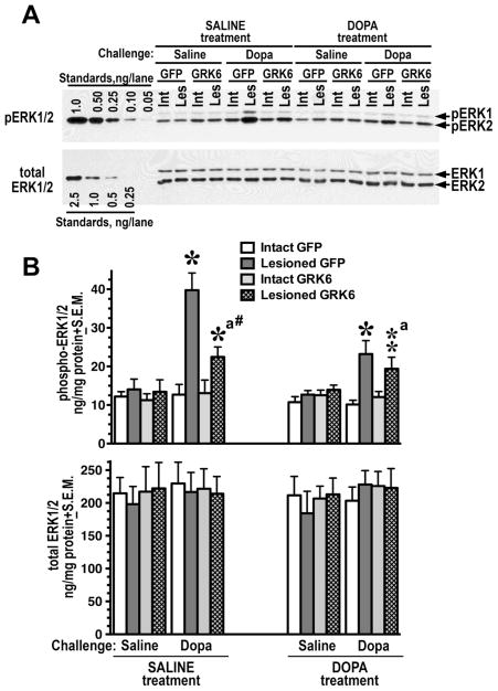
Figure 3. Overexpression of GRK6 in the striatum reduces ERK activation induced by acute L-DOPA.
(A) Western blot showing activation of ERK1/2 in the lesioned striatum of rats expressing GFP (control) or GRK6, chronically treated with saline or L-DOPA and challenged with either saline or L-DOPA (upper panel). Lower blot shows total ERK1/2 expression. (B) Quantification of Western blots of ERK1/2 activation by acute L-DOPA challenge in rats expressing GFP or GRK6 in the lesioned striatum and chronically treated with either saline (drug-naïve) of L-DOPA for 10 days. Rats were challenged with either L-DOPA or saline 45 min prior to sacrifice. N=9–10 rats per group. * - p<0.001 ** - p<0.01, to intact striatum, paired Student’s t-test; a –p<0.05 – to GFP-expressing animals by two-way ANOVA separately in the lesioned striatum in L-DOPA-challenged groups across treatments; # - p<0.05 to the GFP-expressing SALINE-treated L-DOPA-challenged group, lesioned striatum, group, unpaired Student’s post hoc t-test.

