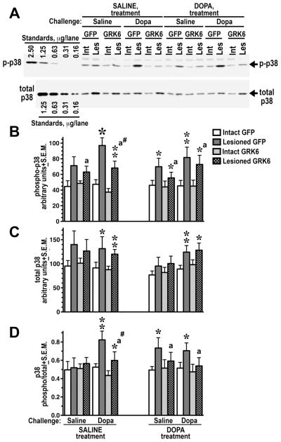
Figure 4. Overexpression of GRK6 in the striatum reduces p38 phosphorylation induced by acute L-DOPA.
(A) Western blots showing activation of p38 in the lesioned striatum of rats expressing GFP (control) or GRK6, chronically treated with saline or L-DOPA, and challenged with either saline or L-DOPA (upper panel). Lower blot shows total p38 expression. (B) Western blots showing activation of p38 in the lesioned striatum of rats expressing GFP (control) or GRK6, chronically treated with saline or L-DOPA and challenged with either saline or L-DOPA. (C) Quantification of the Western blot data for total p38 expression. (D) Ratio of phospho-p38 to total p38 calculated for each animal quantified across experimental groups. N=9–10 rats per group. * - p<0.001, ** - p<0.01, * - p<0.5, to intact striatum, paired Student’s t-test; a – p<0.05 – to GFP-expressing animals by three-way ANOVA separately in the lesioned striatum across treatments and challenges; # - p<0.05 to the GFP-expressing SALINE-treated L-DOPA-challenged group, lesioned striatum, group, unpaired Student’s post hoc t-test.

