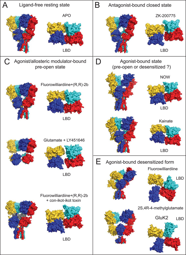
Figure 2. Structures of GluA2 AMPAR and GluK2 kainate receptors in various functional states.
Surface representation of the structures in ligand-free (A), antagonist-bound closed (B), agonist and allosteric modulator-bound pre-open (C) and agonist-bound (D) and agonist-bound desensitized (E) states are shown as viewed from the side for the whole receptor (left) and from top for the LBD tetramer (right). Ligand combinations used and PDB IDs for the structures are shown for each structure (Table 1). Subunits are colored as yellow, cyan, magenta and blue for the subunits A,B,C and D, respectively. The con-ikot-ikot toxin molecule in panel C is colored in grey.

