Fig. 2.
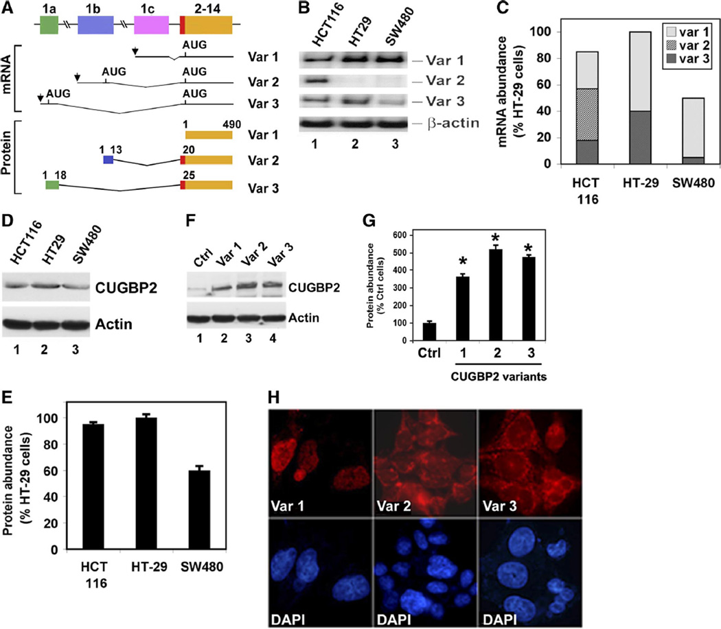
Differential expression of CUGBP2 mRNA in human colorectal cancer cells. A: schematic representation of the origin and structure of the 3 CUGBP2 variants in 3 human cell lines. Like the mouse CUGBP2, alternative promoter usage generates 3 transcript variants in the human cells, resulting in the presence of distinct exon 1 (1a–1c). Human CUGBP2 variant 1 is also 490 amino acids long starting from the AUG in exon 2. Variant 2 is similar to that seen in mouse, with addition of NH2-terminal 19 amino acids. However, variant 3 in the human is not as long as that observed in the mouse, with the addition of only 24 NH2-terminal amino acids. B: RT-PCR analyses from 3 human colon cancer cell lines, HCT116, HT-29, and SW480, show that variant 1 is the predominant isoform in all the cell lines. Moreover, HCT116 has higher levels of expression of all 3 variants. β-actin was used as internal control for loading. C: distribution and abundance of CUGBP2 variant mRNAs in 3 colon cancer cell lines. The percentage was calculated considering CUGBP2 mRNA expression in HT29 as 100%. Data from 3 independent RT-PCR experiments. D: Western blot analysis for CUGBP2 expression in colon cancer cell lines. HT29 has the highest level of protein expression compared with HCT116 and SW480 cells. E: abundance of the CUGBP2 protein. Again, percentage was calculated using expression in HT-29 cells as 100%. Data from 3 independent experiments. F: Western blot analysis for CUGBP2 expression after transient transfection. Plasmids encoding Flag-tagged CUGBP2 variants were transiently transfected in HCT116. Extracts were tested for expression of the transfected proteins. Data shows the higher levels of transfected CUGBP2 variant proteins in the cells. G: abundance of CUGBP2 protein after transient expression of individual isoforms. Variant 1 is least expressed of the 3 variants. Data from 3 independent experiments. *P < 0.05 H: immunocytochemistry analysis for localization of the CUGBP2 variants following transient transfection. Whereas variant 1 is nuclear, variants 2 and 3 are predominantly cytoplasmic. 4′-6-Diamidino-2-phenylindole (DAPI) is used to stain the nucleus.

