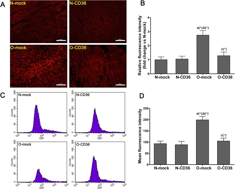
Fig. 6.

Cardiospecific CD36 silencing attenuated abnormal cardiac ROS generation in HFD fed mice. a and b Determination of in situ ROS generation in cardiac tissue by DHE staining. a Representative fluorescence microscopic images of DHE staining (red color) of cardiac frozen sections from each of the four groups (scale bar indicates 100 μm). b Relative fluorescence intensity which stands for levels of ROS generation. c and d Evaluation of ROS generation in isolated ventricular myocytes by DCFH-DA staining. c Isolated ventricular myocytes were incubated with DCFH-DA, and analyzed by flow cytometry. d Mean fluorescence intensity which stands for levels of ROS production. Quantitative and statistical analyses revealed significant increase in cardiac ROS productions in obese mice, but this was reconciled by selectively CD36 silencing. Values are mean ± SD, three pictures of each mouse were analyzed and N = 3 for each group in the DHE staining. A total of 100 000 events were analyzed for each mouse and N = 4 for each group in the DCFH-DA staining. a, vs. N-mock; b, vs. N-CD36; c, vs. O-mock; (*) P < 0.05
