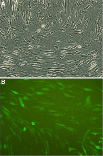
Fig. 2.

hMSC imaging of hMSC infected TGFβ-1 gene with reporter gene GFP observed under fluorescence microscope and inverted microscope. a hMSCs which have been penetrated by the green fluorescent protein (GFP) begin to glow with bright green. Original magnification. b Cells arranged regularly. The shape of gene-transduced hMSC was essentially the same as no-genetically modified cells. Original magnification. Scale bars = 100 μm for (a), 50 μm for (b)
