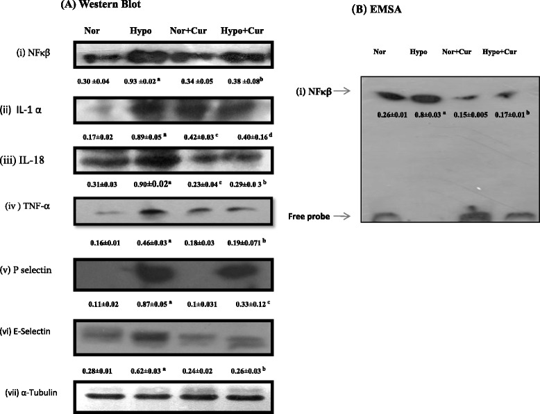
Fig. 2.

Protein expression pattern of NF-κB and related genes in the brain homogenate of rats exposed to hypobaric hypoxia for 24 h at 7620 m. a Representing Western blot analysis of (i) NF-κB, (ii) IL-1, (iii) IL-18, (iv) TNF-α, (v) P-selectin and (vi) E-selectin proteins in the brain of rats under hypoxia. b Depicts the NF-κB-DNA-binding activity. Nuclear extracts were prepared and used to analyse the NF-κB-DNA binding by EMSA. The arrow indicates the position of NF-κB and free probe. The enhanced NF-κB and its downstream genes were downregulated by curcumin treatment prior to hypoxia exposure. The densitometry analyses were represented below their respective analyses. Values are mean ± SD (n = 6). a p < 0.001 compared with normoxia (0 h) group; b p < 0.001 compared with hypoxia (24 h) group; c p < 0.05 compared with normoxia (0 h) group; d p < 0.05 compared with hypoxia (24 h) group. Nor normoxia, Hypo hypoxia, Cur curcumin (100 mg/kg BW)
