
Fig. 9.
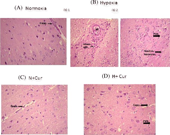
Histological examination of the different groups of rat brain tissues (haematoxylin-eosin staining at 40×). a Photomicrograph from section of the cerebral cortex of the animal under normoxic condition showing normal arrangement and structure of cortical neurons. A cerebral capillary with normal configuration is seen at the upper right corner. b Photomicrograph from section of the cerebral cortex of the animal exposed to 24 h of hypoxia showing numerous inflammatory cells around a blood vessel (BV). Photomicrograph from another area of section of cerebral cortex of animal exposed to 24 h of hypoxic condition showing scattered inflammatory cells (b1) and reactive astrocytes (b2). c Normoxic animals treated with curcumin showed normal configuration in the brain tissue sections. d Photomicrograph from section of the cerebral cortex of the animal exposed to 24 h of hypoxic condition, treated with 100 mg/kg BW of curcumin, showing normal arrangement and structure of cortical neurons without inflammatory cells. A cerebral capillary with normal configuration is seen in the right half (d). Cap capillary structure, BV blood vessels, Inflam cells inflammatory cells, PVS perivascular space, PCE pericellular edema, N normoxia, H hypoxia, Cur curcumin