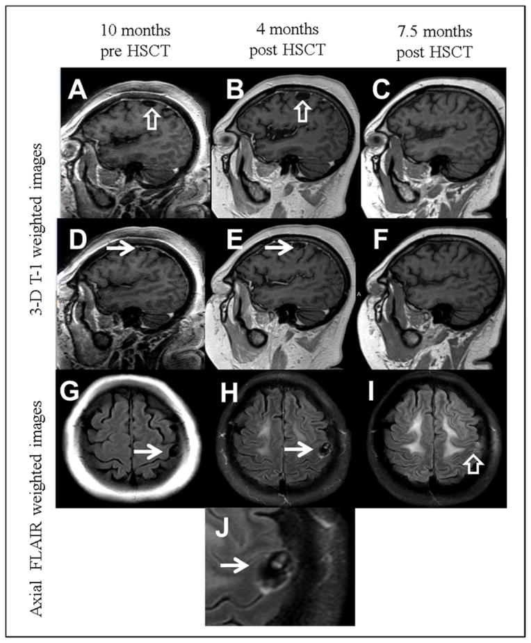
Fig. 1.
Magnetic resonance imaging of the neurocysticercosis lesion. Three-D T1-weighted images of the brain obtained 10 months before hematopoietic stem cell transplantation (HSCT) (A, D), 4 months after transplantation (B, E), and 7.5 months after transplantation (C, F). A small extra-axial cystic structure is identified adjacent to the left precentral gyrus (open white arrows), slightly increasing in size between the first and second examination (A and B). An eccentric hyperintensity within the cyst (solid white arrows) is suggestive of a scolex. The imaging is highly suggestive of subarachnoid neurocysticercosis. The last examination (C and F) show resolution of the cystic structure and internal scolex. (G–J) Axial FLAIR weighted images across the frontal lobes show a cystic structure with fluid signal intensity (white arrows) in G (pretransplant) and H (4 months after transplant). The last examination (I) shows slight residual hyperintensity at the location of the resolved cyst, suggestive of gliosis. (J) is a detail from H showing a clear appearance of a scolex.

