
Figure 1. SD208 inhibits TGFβ signaling in PC-3 cells in vitro and reduces the development of bone metastases from PC-3 in mice.
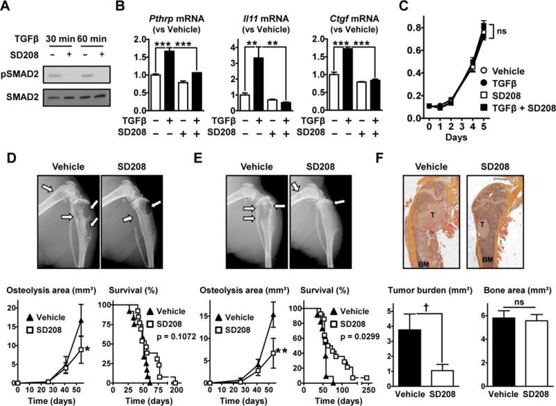
A. Western blot analysis of SMAD2 phosphorylation in PC-3 cells pre-treated ±SD208 (1 μM, 1 hr) and further cultured with TGFβ (5 ng/mL, 30–60 min) before lysis. B. Pthrp, Il11 and Ctgf mRNA measured by RT-qPCR in PC-3 cells cultured ±TGFβ (5 ng/mL) and ±SD208 (1 μM) for 24hr (n = 3). C. Growth of PC-3 cells cultured in the presence or absence of TGFβ (5 ng/mL) and SD208 (1 μM) assessed by MTT assay (n = 6). D,E&F. Nude mice inoculated with PC-3 cells received SD208 (50 mg/kg/day) or its vehicle (D) in a therapeutic manner (starting on day 26) or (E&F) in a preventive manner (starting 3 days before cell inoculation) (n = 11 to 14 mice per group). D&E. (Upper) Representative radiographs (arrows indicate osteolytic lesions). (Lower) Quantification of the osteolysis area on radiographs and Kaplan-Meyer analysis of mouse survival with a log-rank (Mantel-Cox) test. F. (Upper) Representative H&E stained sections of femurs (T, tumor; BM, bone marrow) and (Lower) histomorphometric analysis of tumor burden and bone areas. Results are expressed as the mean ± SEM. ns, not significant, * p < 0.05, ** p < 0.01 and *** p < 0.001 using a 2-way ANOVA with Bonferroni’s posttest. † p < 0.05 using a Student’s t test.