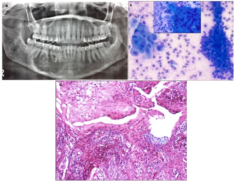
Figure 3.
a) Orthopantomogram shows a large well-defined expansile lytic lesion involving the body of mandible, in bilateral paramidline regions. No cortical breech / periosteal reaction / tooth displacement / root resorption seen. b) Aspirate shows cohesive cluster of basaloid epithelial cells with peripheral palisading and polygonal squamous cells with dense inky blue cytoplasm (Inset) in a proteinaceous background, suggestive of ameloblastoma (MGG x 100). c) Histopathology confirms the squamous differentiation within the basaloid clusters (H&E x200).

