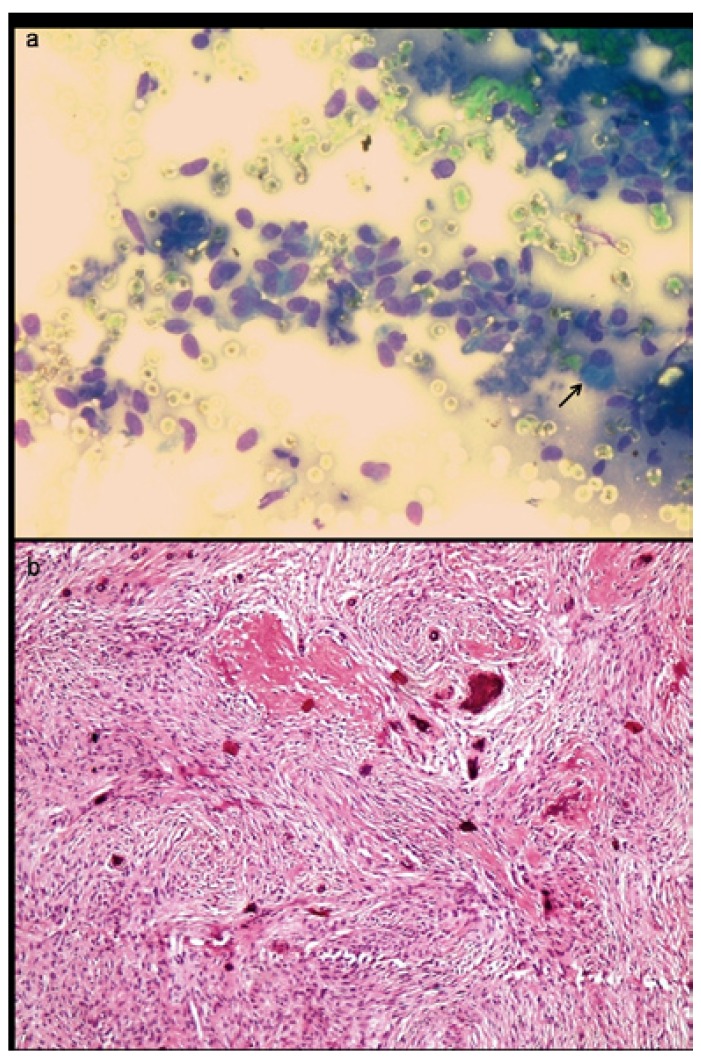
Figure 5.
a) Aspirate smear from a mixed radiolucent lesion in maxilla shows presence of numerous plump fibroblastic cells and occasional osteoblast, suggestive of fibrosseous lesion (MGG x400). b) Histopathology shows predominantly fibroblastic stroma and irregular deposits of pink hyaline cementum like material with central calcification, confirming the diagnosis of cemento-ossifying fibroma (Hematoxylin & Eosin x100).

