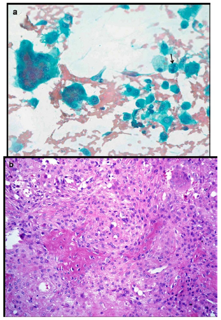
Figure 6.
a) Aspirate smear shows plasmacytoid osteoblasts having moderate amount of cytoplasm with round nuclei, fine chromatin and distinct, single nucleolus. Few benign appearing osteoclastic giant cells and occasional binucleated cells are also seen (Pap x400). b) Histopathology confirms a bone forming tumor comprising osteoid surrounded by rim of epithelioid osteoblasts. Intervening loose fibrovascular stroma has scattered osteoclasts (H&E x200).

