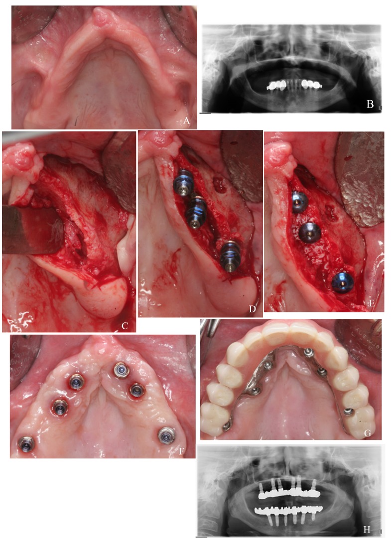
Figure 1.
Test group case: a) preoperative clinical image; b) preoperative panoramic radiograph; c) narrow alveolar ridge of the second quadrant; d) palatal positioned implants in canine and premolar positions; e) particulate bone graft covering exposed threads in the palatal side; f) healed soft tissues; g) placement of the metal-resin screwed prosthesis; h) panoramic radiograph after 5 years of follow-up.

