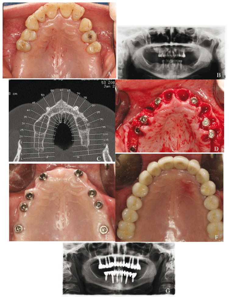
Figure 2.
Control group case: a) preoperative clinical image; b) preoperative panoramic radiograph; c) CBCT scan to study bone availability; d) placement of 8 post-extraction implants, well-centered in the alveolar crest; e) healed soft tissues 1 months after the surgery; f) metal-ceramic fixed prosthesis; g) panoramic radiograph taken after 5 years of follow-up.

