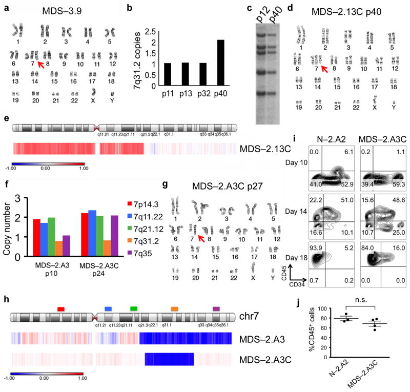
Figure 3. Spontaneous compensation for chromosome 7q dosage imbalance rescues the hematopoietic defect of MDS-iPSCs.
(a) Karyotype of line MDS-3.9, derived from patient no. 3, harboring a derivative chromosome from a 1;7 chromosomal translocation [der(1;7)(q10;p10), the entire long arm of one copy of chromosome 7q is missing and part of chromosome 1q is translocated in its place, identical to the translocation seen in all MDS-iPSC lines from patient no. 3, see also Fig. 1c, MDS-3.1], in addition to two normal chromosomes 7.
(b) qPCR measurement of copy number of a region on 7q31.2 in the del(7q)-iPSC line MDS-2.13 at increasing passage numbers, as indicated.
(c) Southern blot probing integration sites of the vector used for reprogramming of the MDS-2.13 line at passage number 12 (haploid for 7q) and 40 (diploid for 7q).
(d) Karyotyping of MDS-2.13 (see Fig. 1c) at passage number 40 (MDS-2.13C) showing duplication of the normal chromosome 7 without additional karyotypic changes.
(e) aCGH analysis confirming the karyotypic finding. The red color indicates amplification (3 copies) and the white color normal diploid dosage.
(f) qPCR measurement of copy number with different probes along the length of chromosome 7, as indicated, in the del(7q)-iPSC line MDS-2.A3 at passage 10 and 24 (MDS-2.A3C).
(g) Karyotype of line MDS-2.A3C.
(h) aCGH analysis of the del(7q)-iPSC line MDS-2.A3 at passage 10 (MDS-2.A3) and passage 40 (MDS-2.A3C). The blue color indicates deletion (1 copy) and the white color normal diploid dosage.
(i) CD34 and CD45 expression at days 10, 14 and 18 of hematopoietic differentiation. Representative panels of the normal isogenic line N-2.A2 and the dosage corrected MDS line MDS-2.A3C.
(j) CD45 expression at day 14 of hematopoietic differentiation. Mean and SEM are shown. n.s.: not significant.

