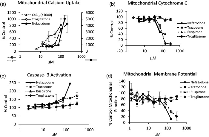
Figure 4.
Steps in the induction of intrinsic apoptosis established through the use of three genetically encoded biosensors and the diffusible probe TMRE in HepG2 cells in static culture. (a) Apoptosis induction resulting from mitochondrial calcium uptake measured by the biosensor Case12-mito (Evrogen). Mitochondrial free calcium is increased at 16 h following addition of CaCl2 in microplates containing Case12-mito transduced HepG2 cells or by addition of the hepatotoxins nefazodone (2nd axis) and troglitazone. (b) Apoptosis arising from mitochondrial damage is monitored at 16 h in HepG2 cell transduced with the cytochrome c-GFP biosensor in response to nefazodone and troglitazone but not the non-hepatotoxins trazodone or buspirone. (c) Apoptosis induction monitored at 16 h by the activation of caspase-3 in HepG2 cells transduced with the Casper-3BG (Evrogen) FPB by nefazodone and troglitazone but not trazodone or buspirone. (d) Mitochondrial membrane potential in HepG2 cells with TMRE (200 nM) for 1 h demonstrates the decrease in mitochondrial membrane potential as a loss of 605 nm fluorescence under increasing nefazodone and troglitazone dosing. Values represent mean and SD of triplicate measurements.

