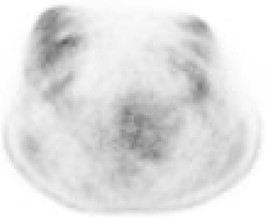
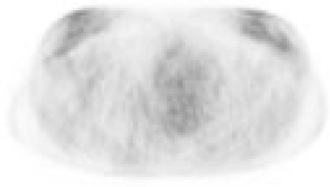
Figure 3.
A 39-year-old patient (Patient C) with a 10 cm low-grade invasive lobular carcinoma of the right breast. 18F-FDG-PET imaging acquired in the (A) prone and (B) supine positions. This tumor exhibited very low metabolic activity and was discordantly categorized as having multifocal distribution on prone scanning but unifocal distribution on supine.


