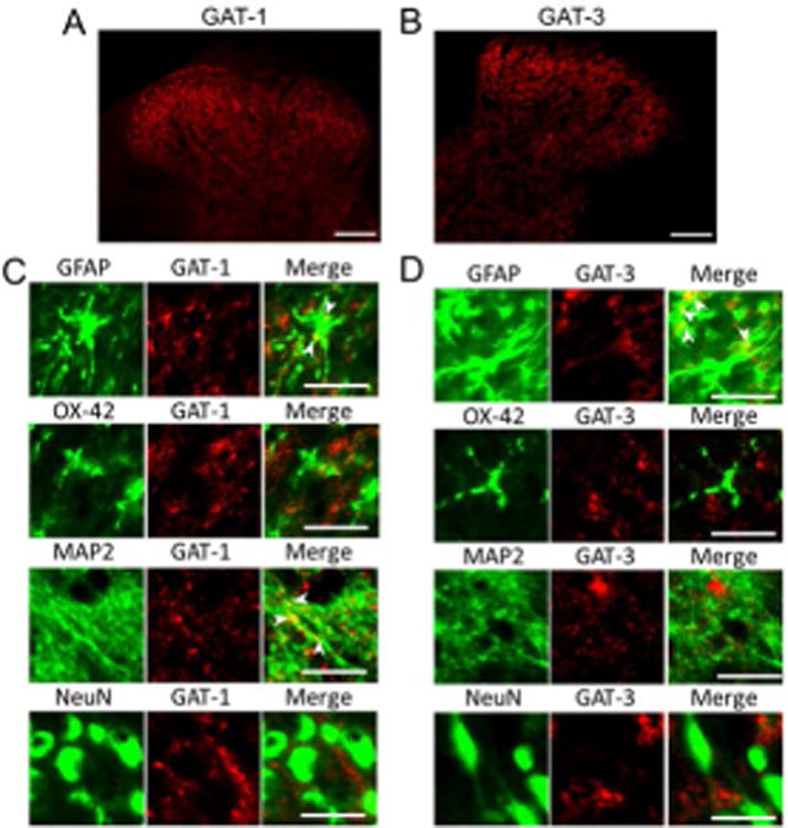
Figure 3. GAT-1 is expressed in neurons and astrocytes and GAT-3 only is present in astrocytes.

Samples were obtained from the spinal dorsal horn of normal control rats. (A) and (B) respectively show staining of GAT-1 and GAT-3 in the spinal dorsal horn, note higher expressions of GAT-1 and GAT-3 at the superficial dorsal horn (Scale bar = 100 μM). (C) Shows that expression of GAT-1 is colocalized with MAP2 (a marker for neuronal cytoskeleton) and GFAP (an astrocyte marker), but not with NeuN (a neuronal cell body marker) or OX42 (a microglia marker) (Scale bar = 20 μM). (D) Shows that GAT-3 is predominantly colocalized with GFAP, but not OX42, MAP2, or NeuN (Scale bar = 20 μM).
