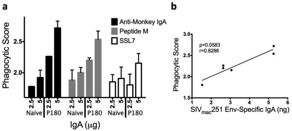
Fig. 8. ADCP activity of IgA purified by several methods from feces.
THP-1 cells were incubated with SIVmac251 gp120-coated fluorescent beads and IgA from feces. After 3 hours, phagocytosis was quantified by flow cytometry. (a) Fecal IgAfrom a naïve monkey and an infected monkey (P180) were purified using α-Mon IgA, Peptide M, and SSL7 columns, respectively, and IgA was used in the ADCP assay. (b) The phagocytic score was plotted against the amount of SIVmac251 Env-specific IgA present in each P180 sample used.

