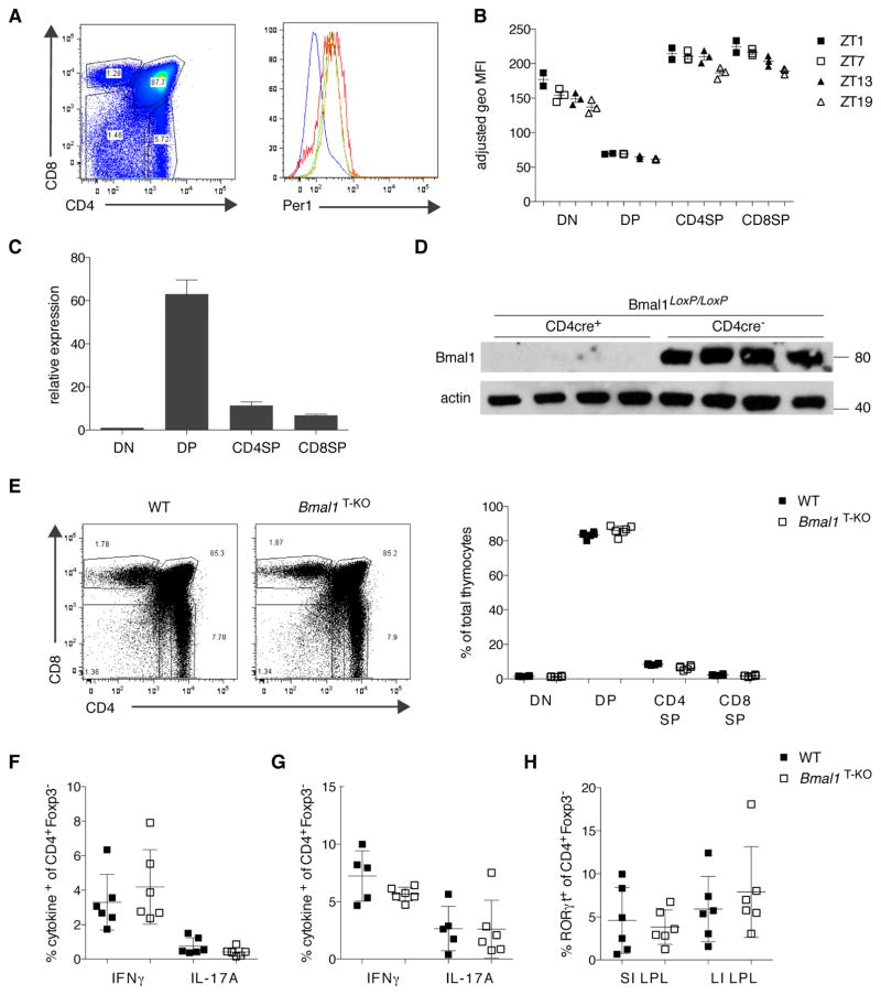
Figure 1. Dynamic Regulation of Circadian Reporter Expression During Lymphocyte Development but T Cell-Specific Deletion of Bmal1 has no Effect on T Cell Development.
(A) PER1Venus expression in thymocytes was analyzed by flow cytometry. The gated populations in the left plot correspond to the histogram overlay in the right plot (DN: red line; DP: blue line; CD4SP: green line; CD8SP: orange line).
(B) Data as in (A) plotted for individual mice (n= 2–3 animals per time point) analyzed at four time points (ZT1, ZT7, ZT13, ZT19).
(C) Bmal1 expression analysis in FACS purified thymocyte populations from three individual mice. Expression is shown relative to the DN population.
(D) BMAL1 protein levels were assessed by Western blotting in lysates from total thymus of WT or Bmal1T-KO mice. Equal loading was confirmed by β-actin expression.
(E) Thymocyte development is unperturbed in Bmal1T-KO mice.
(F, G) IFNγ and IL-17A production in CD4+ T cells after αCD3/αCD28 stimulation in spleen (F) and large intestine LPL (G) is not affected by BMAL1-deficiency.
(H) Frequencies of RORγt expressing CD4+ T cells in small and large intestine LPL are unchanged in Bmal1T-KO mice.
(B) Error bars represent mean ± SEM. (C, E–H) Error bars represent mean ± SD. All data are representative of at least three individual experiments. See also Figure S1, 3.

