
Fig. 4.
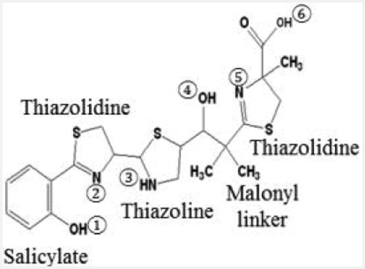
Structure of the Ybt siderophore. Salicylate, thiazolidine, and thiazoline rings as well as the malonyl linker are labelled. The six coordinate binding sites for Fe3+ are shown.
Official websites use .gov
A
.gov website belongs to an official
government organization in the United States.
Secure .gov websites use HTTPS
A lock (
) or https:// means you've safely
connected to the .gov website. Share sensitive
information only on official, secure websites.
Structure of the Ybt siderophore. Salicylate, thiazolidine, and thiazoline rings as well as the malonyl linker are labelled. The six coordinate binding sites for Fe3+ are shown.