Figure 3.

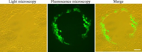
Light microscopy and fluorescent microscopy observations of rRGV-infected GSTC cells. Infected GSTC cells not only showed CPE, but also emitted a green fluorescence signal. Bar = 100 μm.

Light microscopy and fluorescent microscopy observations of rRGV-infected GSTC cells. Infected GSTC cells not only showed CPE, but also emitted a green fluorescence signal. Bar = 100 μm.