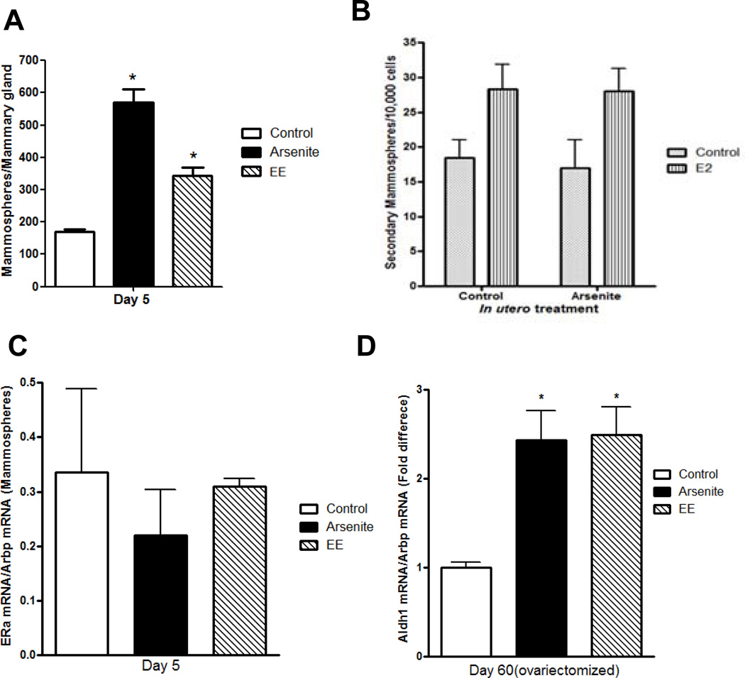
Figure 3. Effects of early life exposure to arsenite on mammosphere-forming cells in the mammary gland of female offspring.
Pregnant female rats were treated with arsenite or ethinyl estradiol as described in Figure 1.
(a) In utero effects on the number of mammospheres obtained from female offspring on postnatal day 5 (mean±SEM; n=4 animals/group; * p<0.05).
(b) In utero effects on estradiol induced proliferation of mammospheres. First generation mammospheres from control animals and arsenite exposed animals were digested with trypsin, stem/progenitor cells were selected in serum-free media under non-adherent conditions in the presence or absence of estradiol (1 nM), and the second generation mammospheres were counted (mean±SEM; n= 2/group).
(c) In utero effects on ERα expression in mammospheres obtained from female offspring on postnatal day 5. ERα mRNA was determined by a qRT-PCR assay and normalized to Arbp mRNA (mean±SEM; n=2/group).
(d) In utero effects on Aldh1 expression in the adult gland. Female offspring were ovariectomized on postnatal day 45, and on postnatal day 60, the amount of Aldh1 mRNA was measured by a qRT-PCR assay, normalized to Arbp mRNA, and presented as fold difference (mean±SEM; n=4–11/group; * p<0.05).

