
Fig. 2.
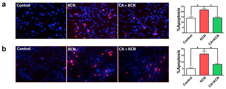
Protective effect of CA on KCN-induced apoptotic cell death in rat and human cortical neuronal cultures. (a) Representative images of primary rat cortical neurons: vehicle-treated control and KCN-exposed with or without treatment with CA. Apoptotic cell death was identified by TUNEL staining (red); all cell nuclei were stained with DAPI (blue). Histogram shows the percentage of apoptotic neurons in each group. (b) KCN-induced apoptotic death and rescue by CA was investigated in human iPSC-derived cortical neurons. All data are expressed as mean + SEM (n = 20; *p < 0.001 by one-way ANOVA with Tukey’s post test).