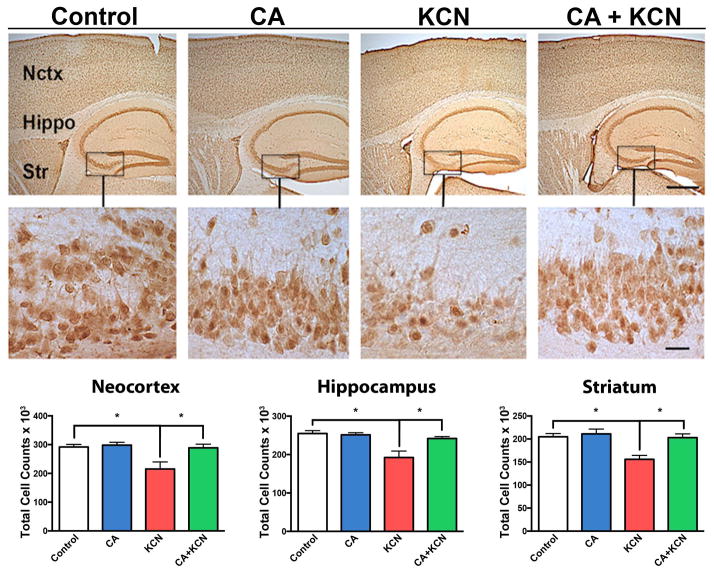
Fig. 6.
Protective effect of CA on KCN-induced loss of neurons. Representative NeuN staining of neocortex (Nctx), hippocampus (Hippo), and striatum (Str) of mouse brain exposed to vehicle control, CA-only, KCN, or CA+KCN. Low and high magnification images are shown on top and bottom, respectively (scale bars, 250 μm at low power and 40 μm at high power). Histograms below show stereological cell counts of the affected brain areas in neocortex, hippocampus, and striatum under different treatment conditions. All data are expressed as mean + SEM (total n = 37; *p < 0.01 by one-way ANOVA with Tukey’s post test).

