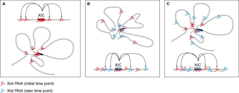
Figure 1.

X chromosome topology directs Xist localization.
(A) Xist RNA (red) utilizes 3D contacts to localize to distal sites. (B) Model 1: the Xic samples the entire X over time and Xist RNA (red and blue at different time points) accumulation is determined by the affinity of Xist RNA for chromatin features at each region. (C) Model 2: Xist RNA (red) alters contact sites such that they have greater affinity for subsequently produced Xist RNA (blue), promoting its spread in cis along the X.
