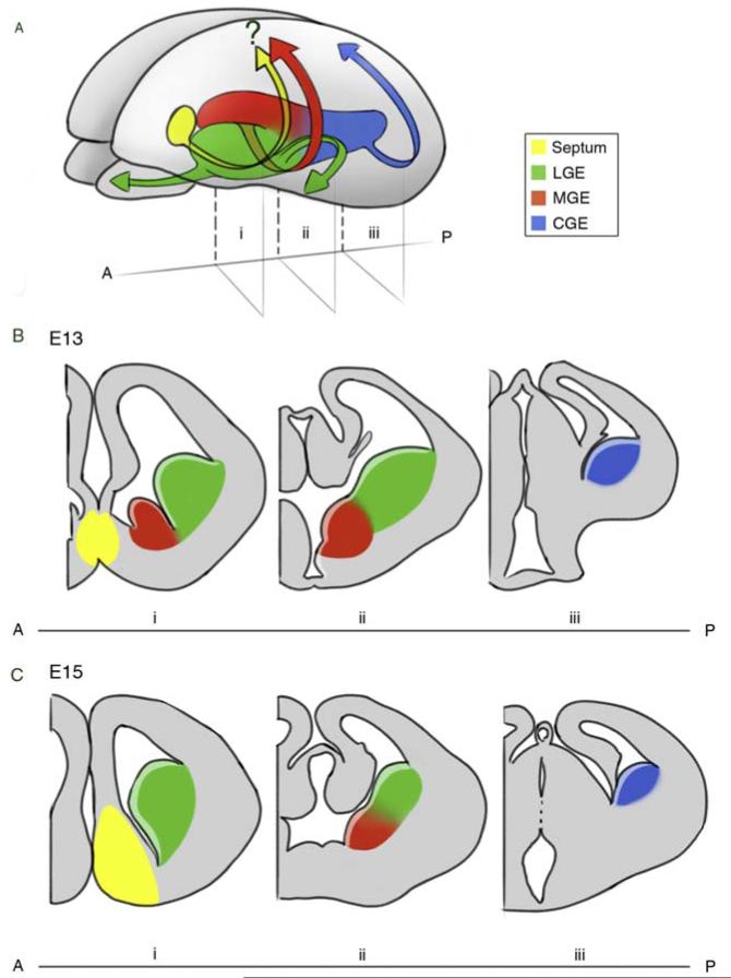
Figure 3.1.
Schematic representation of the developing murine brain. (A) Three-dimensional view of a developing brain indicating the ventral anatomical regions that give rise to interneurons and their correspondent migratory pathways. The LGE (green) generates interneurons that migrate to the olfactory bulb and striatum. Both the MGE (red) and CGE (blue) produce cortical interneurons. Whether the septum produces cortical interneurons (yellow) is still a matter of debate. (B–C) Coronal views of the brain in (A) at three locations (i–iii) along the anterior (A)–posterior (P) axis at the embryonic ages E13 (B) and E15 (C).

