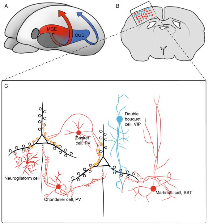
Figure 3.2.
Differential origin of cortical interneuron subtypes. (A) Three-dimensional view of an embryonic murine brain highlighting the two ventral regions that produce cortical interneurons, the MGE (red) and the CGE (blue). (B) Coronal view of an adult brain, illustrating the proportion and relative distribution of cortical interneurons derived from the MGE (red dots) and the CGE (blue dots). (C) Detailed schematic view of the boxed region in (B) illustrating the main interneuron subtypes derived from the MGE (red cells) and CGE (blue cells), and how they characteristically interact with the pyramidal cells (black cells). This figure is adapted from Kawaguchi and Kubota (2002).

