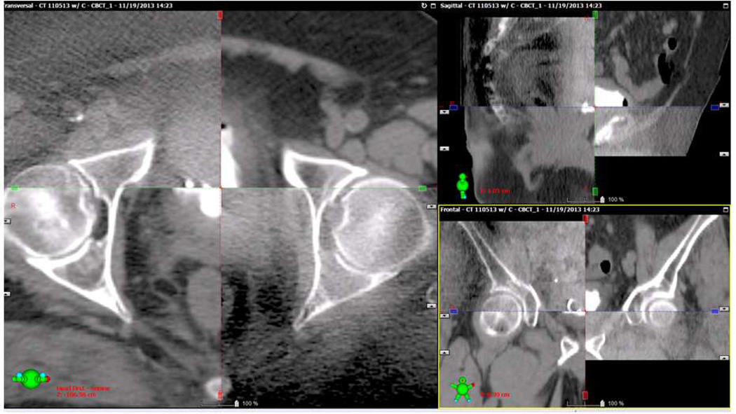
Figure 4. Cone beam CT fused with a treatment planning image.
A cone beam CT was obtained on a patient receiving TMT to verify alignment prior to radiation treatment. In each panel, the cone beam CT image is in the upper left and bottom right portion of the image, while the planning CT image is in the upper right and lower left portion. Note that cone beam CT image quality is inferior to the diagnostic image. Overlay of the images allows comparison of the anatomy between scans to verify alignment of bony and soft tissue structures. Transverse, sagittal, and coronal reconstructions are presented.

