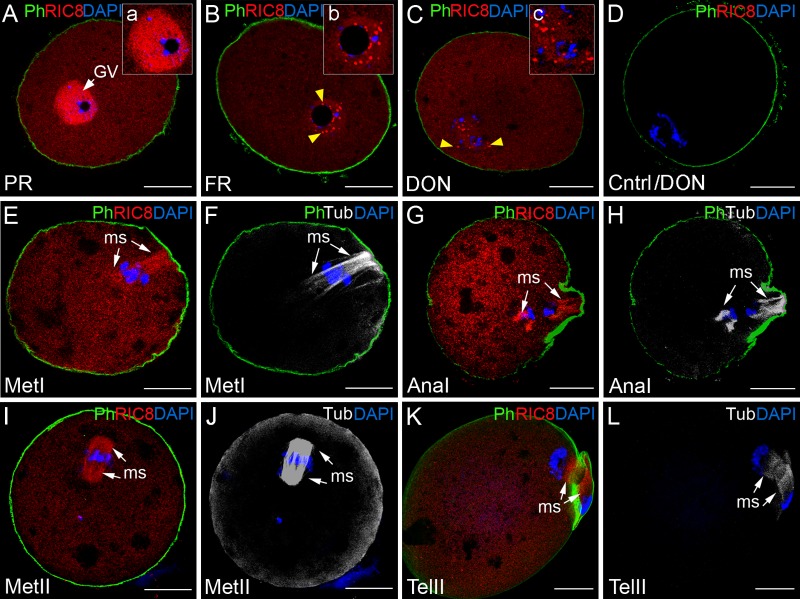
Fig 2. Dynamic changes in localization of RIC8 in the mouse oocyte during meiosis.
RIC8 was visualized with RIC8 antibody (red), microtubules with antibody to β–tubulin (Tub, white), chromatin with DAPI (blue) and F-actin with phalloidin (Ph, green). (A, B) GV- stage oocyte; (C, D) GVBD (germinal vesicle breakdown) stage oocytes; (E-H) oocytes undergoing meiosis I or (I-L) meiosis II. (a`, b`, c`) Higher magnification of the region of chromatin. Yellow arrowheads indicate the RIC8 foci at nucleolus and on chromatin. White arrows point to the meiotic spindle. (D) Negative control. Abbreviations: AnaI, anaphase of meiosis I; DON, disappearance of the nucleolus; GV, germinal vesicle; FR, full rim stage; MetI/MetII, metaphase of meiosis I or II; ms, meiotic spindle; PR, partial rim stage; TelII, telophase of meiosis II. Scale bar: 20 μm.

