
Figure 2.
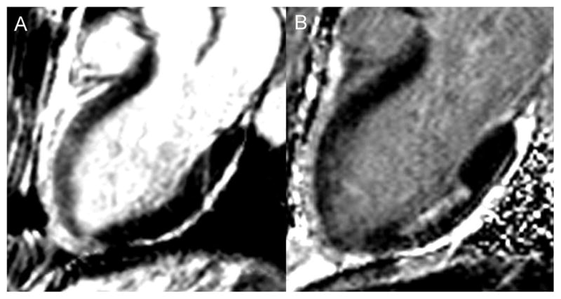
A – 3-chamber long axis bright blood T2-W image in a patient with a spontaneous LAD dissection demonstrating bright signal in the anterior wall consistent with edema. B – 3-chamber long axis phase sensitive inversion recovery late gadolinium enhanced image in the same patient showing absence of LGE in the anterior wall, but evidence of a prior 50% transmural infarct in the mid inferior wall. Together these images demonstrate evidence of myocardial stunning without infarction in the anterior wall.