
Fig. 3.
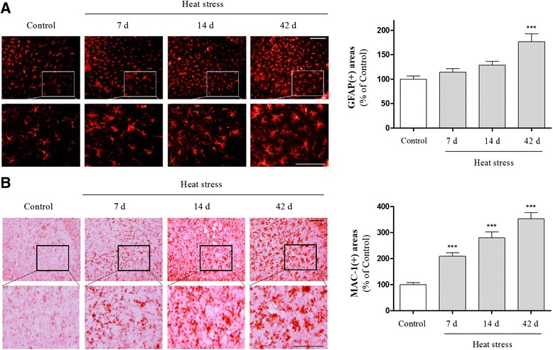
Time-course analysis of heat stress-induced activation of astrocytes and microglia in mouse hippocampus. The presence of astrogliosis (a) and microgliosis (b) was determined using glial fibrillary acidic protein (GFAP) and macrophage-1 antigen (Mac-1) staining, respectively. Quantification of GFAP- and Mac-1-stained cells was performed by measuring the area fraction of GFAP and Mac-1-immunoreactive cells/areas in the CA3 of the hippocampus. Scale bar = 50 μm. Values are expressed as means ± S.E.M. *** p < 0.001 indicates that the mean value was significantly different from the control group