
Fig. 4.
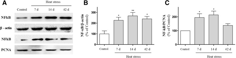
Effects of heat exposure on the nuclear factor (NF)-κB expression in hippocampal whole-tissue lysates and nuclear extracts as measured by Western blot analysis. (a) Representative Western blot illustrating the expression of NF-κB in the hippocampus. The graphs display densitometric analyses of the expression ratios of NF-κB/β-actin in whole protein extracts (b) and of NF-κB/ proliferating cell nuclear antigen (PCNA) in nuclear extracts (c) from the hippocampus. Values are expressed as means ± S.E.M. *p < 0.05 and **p < 0.01 as compared with the control group