
Fig. 7.
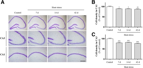
Effects of heat stress on cell death in the hippocampus. (a) Representative pictures of Cresyl violet staining in the granule cell layer and the pyramidal cell layer of the hippocampus. Cresyl violet-stained cells were markedly lower in the heat-treated groups than the control group. The number of cells was reduced significantly in both the CA1 (b) and CA3 (c) regions. Values are expressed as means ± S.E.M. *p < 0.05, **p < 0.01, and ***p < 0.001 as compared with the control group