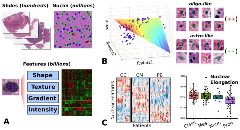
Figure 3.
Quantitative nuclear morphometry. (A) Image analysis algorithms are used to delineate nuclei in whole-slide images. A set of features is calculated to describe the appearance of each nucleus. This system is capable of processing thousands of slides and hundreds of millions of nuclei. (B) We developed a model-based system to score nuclei based on oligodendroglial differentiation. This model was validated by correlation of nuclear scores and gene expression data. (C) Model-free approaches were used to explore the clinical and genomic associations of nuclear features. Clustering of patient morphological signatures revealed three distinct patient clusters. Unsupervised analysis of features shows that proneural tumors are associated with more round, regular nuclei.

