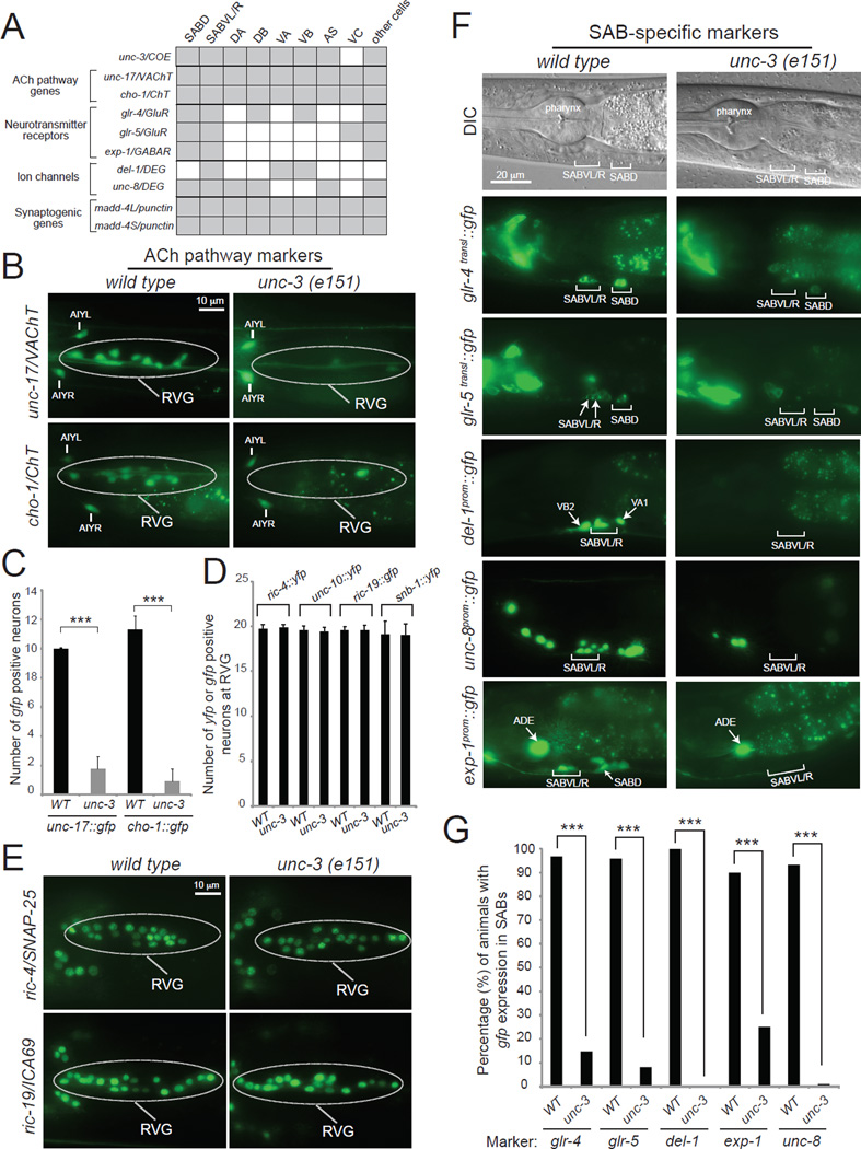
Figure 4. unc-3 is required for multiple aspects of neuronal signaling in the SAB motor neurons.
A: A combinatorial code of gene expression for SAB head MNs based on our findings and WormBase (www.wormbase.org). Grey shaded boxes indicate expression in the respective neuron type; white boxes indicate absence of expression.
B, C: ACh pathway genes (unc-17/VAChT, cho-1/ChT) fail to be expressed at the retrovesicular ganglion (RVG) of unc-3(e151) mutants. The cell bodies of SABs are located at the RVG together with other (around 7–9) cholinergic neurons. Scale bar 10 µm. Error bars represent standard deviation (s.d). *** : p value < 0.001. N > 10 for unc-17prom::gfp in WT and unc-3(e151) animals. N = 22 for cho-1fosmid::gfp in WT and unc-3(e151) animals. Animals at the first day of adulthood were analyzed.
D, E: Transcriptional reporters for panneuronally-expressed genes (ric-4, ric-19, unc-10, and snb-1) are unaffected in unc-3(e151) mutants. The cell bodies of SABs are located at the RVG together with other 17 neurons. Scale bar 10 µm. Detailed Quantification provided in D. Error bars represent standard deviation (s.d). N > 10 for ric-4fosmid::yfp, ric-19prom::gfp, unc-10fosmid::YFP, and snb-1fosmid::YFP in WT and unc-3(e151) animals. Animals at the first day of adulthood were analyzed. See also Figure S1.
F, G: The expression of neurotransmitter receptors (glr-4, glr-5, exp-1) and ion channel-encoding genes (del-1, unc-8) is significantly affected in the SAB neurons of unc-3(e151) mutants. See also Figures S1 and S4. Scale bar 20 µm. Quantification provided in G. N = 25 for glr-4, glr-5, del-1, and unc-8 reporters in WT and unc-3(e151) animals. N = 20 for exp-1 reporter in WT and unc-3(e151) animals. Animals at the fourth larval (L4) stage were analyzed. In the quantification graph (G), the percentage of animals expressing the reporter in SABVL/R and SABD is shown for glr-4 and glr-5, while for del-1 and unc-8 the percentage of animals expressing the reporter in SABVL/R is shown. Similar results were obtained when a fosmid-based reporter was used to monitor UNC-8 protein expression in the SAB neurons (100 % of wdEx948 [unc-8fosmid::gfp] animals (N=35) showed gfp expression in the SABV neurons, while 42.85 % of unc-3(e151); wdEx948 [unc-8fosmid::gfp] showed gfp expression in SABV neurons (N=35). For exp-1, the percentage of animals with expression in SABD is shown. *** : p value < 0.0001.

