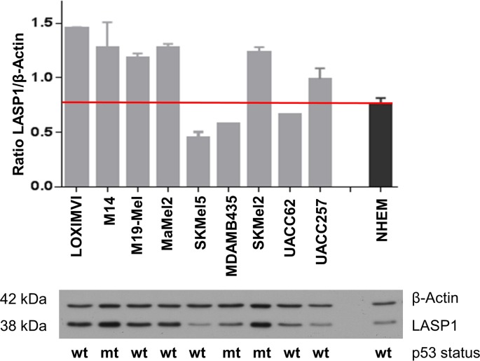
Fig 2. LASP1 expression in melanoma cell lines.
Expression of LASP1 and β-actin in melanoma cell lines and in normal human epidermal melanocytes (NHEMs) was determined by Western blotting (10 μg protein per lane). Bar graphs represent mean and SEM integrated optical density of Western blot bands from three individual experiments. The mutational status of the p53 oncogene is indicated for each cell line (wt wildtype; mt, mutant).

