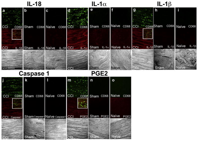
Fig. 4.
Double immunofluorescence for macrophages and cytokines on ipsilateral sciatic nerve cryosections suggests inflammasome activation in CCI rats: IL-18 a shows infiltration of CD68 positive macrophages and presence of IL-18 in CCI sciatic nerve. Sham and naïve control sciatic nerves did not exhibit presence of macrophages and IL-18 (b, c). IL-1α ipsilateral CCI sciatic nerves exhibit presence of macrophages and IL-1α (D), which are both absent from sham and naïve control tissue (e, f). IL-1β macrophages and IL-1β are evident in the affected sciatic nerve (g), but are absent from sham and naïve control tissue (h, i). Caspase-1 macrophages and caspase-1 are evident in the CCI sciatic nerve (j), but are not evident in the sham and naïve control tissue (k, l). PGE2 macrophages and PGE2 are evident in the affected CCI sciatic nerve (m). No macrophages or PGE2 expression are evident in the sham and naïve control tissue (n, o). For each experiment group (a, b, c), (d, e, f), (g, h, i), (j, k, l) and (m, n, o), the confocal images were acquired in the same sitting with the same image acquisition parameters. Bar = 150 μm.

