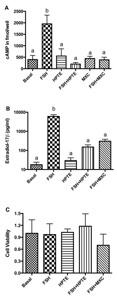
Figure 1. Effect of MXC and HPTE on FSH-stimulated intracellular cAMP and estradiol-17β (E2) production and cell viability.
Granulosa cells were incubated in the absence (basal) or presence of FSH (3ng/ml), HPTE (10 μM), a combination of HPTE and FSH, or MXC (10 μM) alone or in combination with FSH for 48 h. Media were collected for E2 radioimmunoassay, and cells were harvested for intracellular cAMP determination or cell viability analysis; these techniques were performed as described in sections 2.4 through 2.6. (A) cAMP was expressed as mean fmol/well ± SEM (n ≥ 3). (B) E2 was expressed as pg/ml ± SEM of culture medium (n = 5). (C) Cell viability measured with the WST-1 assay showed no difference between control and treatments (n = 3). b = significantly different (p < 0.01) from control as determined by one-way ANOVA followed by Dunnett’s multiple comparison post-hoc analysis.

