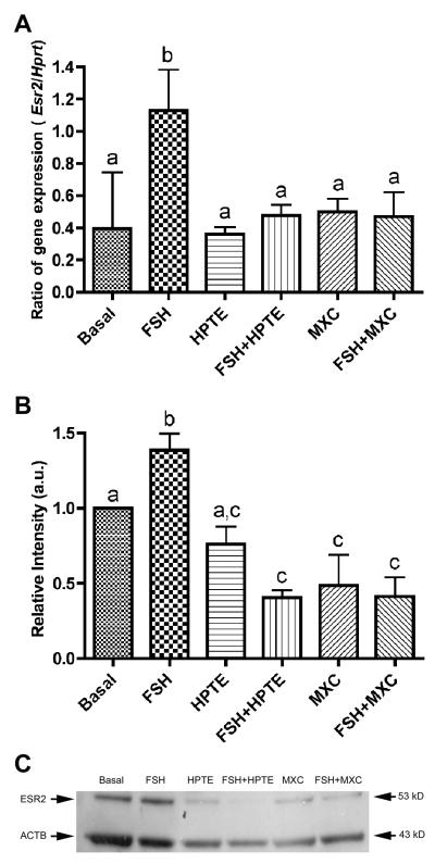
Figure 3. Effect of HPTE and MXC on estrogen receptor β (Esr2) mRNA and protein levels in granulosa cells.
(A) Esr2 mRNA was induced by 3 ng/ml FSH-treatment at least 2-fold over basal levels. HPTE (10 μM) or MXC (10 μM) inhibited FSH-induced Esr2 expression. Total RNA was extracted from granulosa cells and analyzed by qPCR for Esr2 mRNA expression as described in section 2.7. All expression values were normalized with Hprt as a reference gene (n = 4). (B) Western blots supported the qPCR result and showed that FSH stimulated the protein levels of ESR2, which were inhibited by HPTE and MXC. ImageJ (NIH) was used to quantify protein band intensity. β-actin (ACTB) was used to normalize ESR2 levels (n = 3) and samples were compared to basal. Different letter indicates significant difference between treatments groups as determined by one-way ANOVA (p < 0.01) followed by Newman-Keul multiple comparison post-hoc analysis. (C) A representative Western blot is shown.

