Fig. 4.
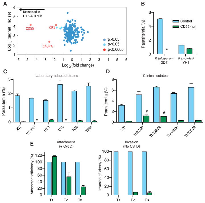
CD55 is a critical host receptor for P. falciparum. (A) Scatter plot of plasma membrane proteins identified by PMP of CD55-null erythrocytes relative to controls from two unrelated individuals, and quantified by > 2 peptides. (B) Invasion of control erythrocytes (blue) or CD55-null erythrocytes (green) by P. falciparum strain 3D7 or P. knowlesi strain YH1. *, below detection. (C) Invasion by laboratory-adapted P. falciparum strains. (D) Invasion by P. falciparum clinical isolates. #, thin smears showed 0.6–1% gametocytes. For B-D: Mean ± SD, N=3. 10,000 cells scored per well by flow cytometry. (E) Efficiency of P. falciparum 3D7 merozoite attachment to the surface of CD55-null (green) versus control (blue) RBCs using cytochalasin D (Cyt D). Invasion was measured in the absence of Cyt D. T1=30 min, T2=60 min, T3=180 min after addition of schizonts (Fig. S10). Attachment to controls at T1 was 3.6–5.3%. Mean ± SD, N=2. 20,000 cells scored per well by flow cytometry.

