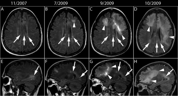
Fig 1.
MRI evolution of MS and PML lesions. (A-D) Axial and (E-H) corresponding sagittal FLAIR images. (A, E) After one year of natalizumab monotherapy, MS lesions are present in the left periventricular area and the splenium of the corpus callosum (arrows). (B, F) Twenty months later a new left subcortical lesion (arrowhead) can be seen in the left frontal lobe, remote from the old MS lesions (arrows). (C, G) Two and a half month later, the left frontal lobe lesion has extended posteriorly and contralaterally through the genu of the corpus callosum to the right frontal lobe white matter (arrowheads), while the MS lesions stable appearance (arrows). (D, H) One month later the PML lesions have spread further in the right frontal lobe and the left frontal and parietal lobes, close, but separate from the MS lesions (arrows).

