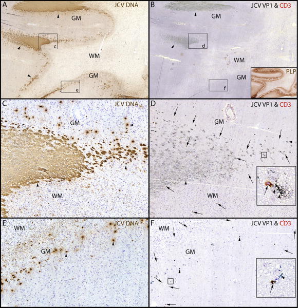
Fig 2.
PML lesions are sharply demarcated and harbor CD3+ T cells.
(A) ISH for JCV DNA (Cresyl violet counterstaining and (B) IHC for JCV VP1 & CD3 show that JCV-infected cells (arrowheads) are mostly restricted to the junction between white matter (WM) and gray matter (GM) and the lower layers of the cortex. Conversely, the deep WM is almost free of JCV-infected cells and totally demyelinated as shown by proteolipid protein staining (PLP, brown (B inset)), but not cavitary. (C-F). Magnifications of rectangles c and e from A show a sharp demarcation of the areas containing JCV-infected cells (arrowheads) at the gray-white junction (C). The edge of JCV infection spreads in the lower layer of the cortex (E, arrowheads). Magnification of rectangles d and f from B, show that cells expressing JCV VP1 protein (arrowheads) are surrounded by CD3+ T cells (marked by arrows in D and F). The majority of these T cells (D and F insets, arrow) are located in close proximity to the JCV-infected cells (D and F insets, arrowheads). Overall, the T cells are mostly found in areas where the cell nuclei contain JCV DNA (C) and express JCV VP1 (D), .

