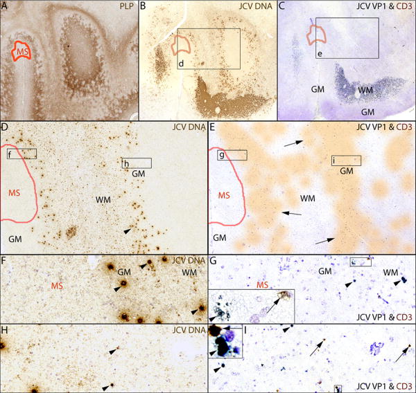
Fig 3.
Proximity of MS and PML lesions in the left occipital lobe
(A-C) IHC staining for PLP (A) shows a subpial MS lesion (delineated in red) and demyelination lesions of PML involving the subcortical white matter (WM) and lower layers of the cortical gray matter (GM), which contain numerous cells harboring JCV DNA (B, JCV ISH & Cresyl violet counterstaining) and expressing JCV VP1 protein in C (LFB counterstaining). (D-E) The magnifications of the boxes s d & e from B & C show that cells harboring JCV DNA (D, arrowheads), those expressing JCV VP1 and CD3+ T cells (E) are not present in the MS lesion. (E) The distribution of the CD3+ T cells is shaded in orange (arrows). (F-I) Higher magnifications of the MS-PML interface (boxes f, g) and the lower GM side of the PML lesion (boxes h, i) show that cells harboring JCV DNA (F, arrowhead), those expressing JCV VP1 (G, arrowheads) and CD3+ T cells (G, arrows) are present at the border of the MS lesion. JCV infection of the gray matter is shown in (H, I arrowheads). Insets in G & I at higher magnification show CD3+ T cells in close proximity of cells expressing JCV VP1 protein.

