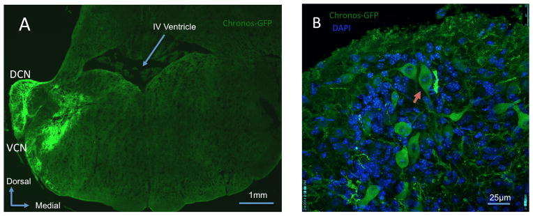
Figure 2. Chronos expression in the cochlear nucleus.

A: Mosaic confocal 63x image showing Chronos-GFP expression within the dorsal cochlear nucleus (DCN) and ventral cochlear nucleus (VCN). B: Confocal 63x image of the DCN demonstrates Chronos-GFP expression within a fusiform cell (red arrow) and in other neuronal and non-neuronal populations. DAPI demonstrates all cell nuclei.
