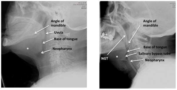
Figure 2. Relevant anatomy seen on lateral film in laryngectomy patient without and without NGT.

Left panel demonstrates proximity of neopharynx to spinal cord in a patient without NGT. Right panel depicts a different patient with NGT coursing though neopharynx. * Depicts likely former location of hyoid bone prior to laryngectomy which is notably absent.
