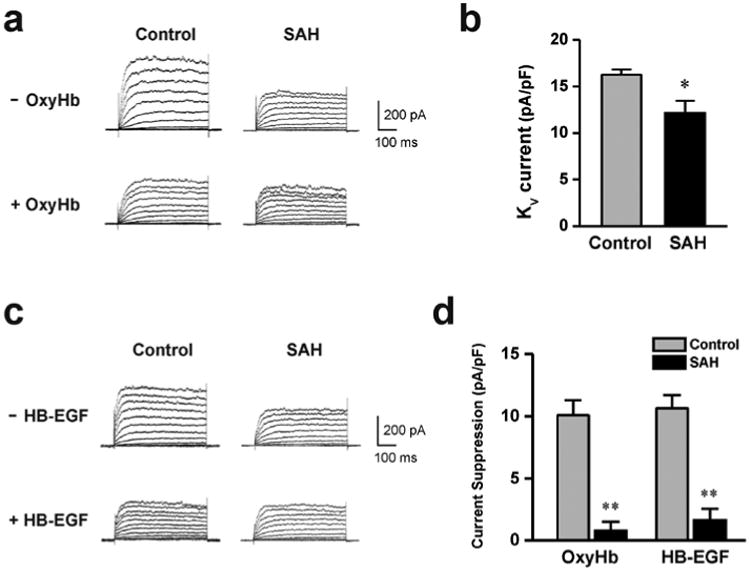
Figure 1. OxyHb and HB-EGF suppressed KV currents of cerebral artery myocytes from control, but not SAH model animals.

a) Example of whole-cell K+ currents before and after 10 minute application of OxyHb (10 μM) to cerebral artery myocytes isolated from a control and SAH model animal. b) Summary of 4-AP-sensitive KV currents obtained from control (n = 6) and SAH model (n= 4) animals. c) Examples of whole cell K+ currents before and after 10 minute application of HB-EGF (30 ng/ml) to cerebral artery myocytes isolated from control and SAH model animals. d) Summary data demonstrating that OxyHb and HB-EGF significantly suppressed KV currents in cerebral artery myocytes from control, but not SAH model animals. OxyHb treatment; control: n = 7, SAH: n = 4, HB-EGF treatment; control: n = 6, SAH: n = 5. ** P < 0.01 vs control, unpaired students t-test.
