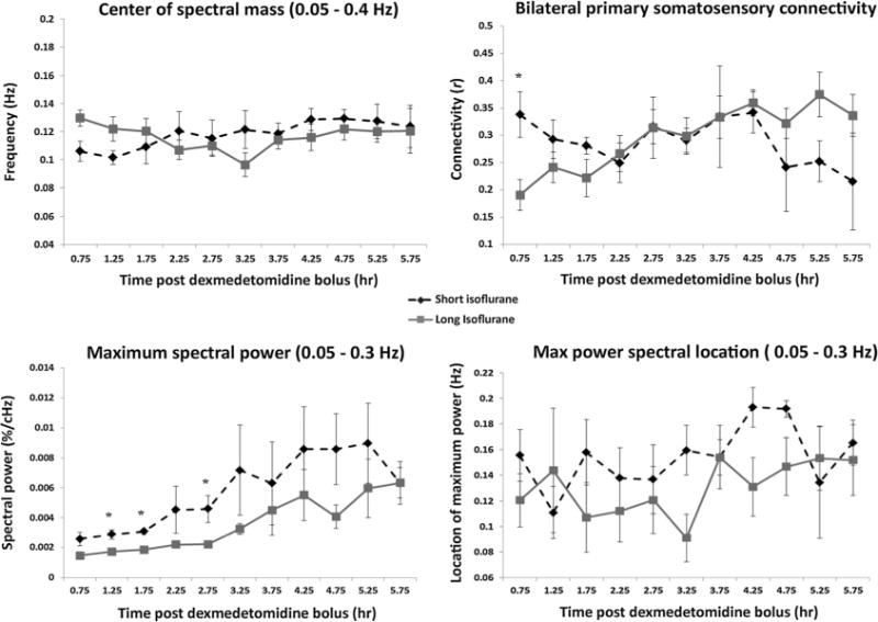
Figure 4.

Spectral characteristic and connectivity evolution. Average group values and SEM are plotted for center of spectral mass (top left), bilateral S1 connectivity (top right), maximum spectral power (bottom left), and the location of that maximum power (bottom right) for both the short (dotted line) and long isoflurane groups (solid line). No significant differences are found between the two groups for spectral CoM or for location of the maximum spectral power. Bilateral functional connectivity exhibits a significant difference between the groups at the 0.75 h time point followed by a convergence in connectivity data at the 2.25 h time point. Significant differences between maximum spectral powers are found at the 1.25, 1.75, and 2.75 h time points. Intra-group evaluations highlighting changes between early data and late data are calculated and presented in Table 2.
