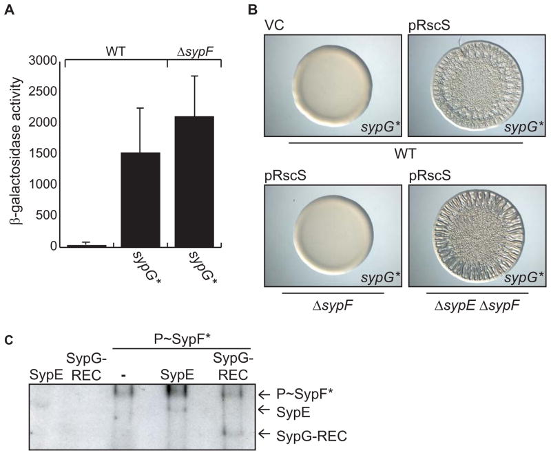
Figure 4. Determining where SypF functions in the Syp pathway.
(A) SypG*-induced PsypA-lacZ reporter activity in WT (KV7230) or ΔsypF (KV7231) strains. Error bars represent standard deviation. (B) Wrinkled colonies of WT V. fischeri strains producing SypG* (KV6527) with vector control (VC, pKV282) or pRscS (pARM7) (top two panels) and of pRscS, SypG*-producing ΔsypF (KV6526) and ΔsypE ΔsypF (KV6586) strains (bottom two panels). Cultures were spotted and colony morphology was assessed after 19 hours. (C) in vitro phosphotransfer assay. Left two lanes: GST-SypE or MBP-SypG-REC incubated with radiolabelled ATP. Right three lanes: phospho-SypF* incubated with or without GST-SypE or MBP-SypG-REC.

